Reduce Your
EGD Uncertainty
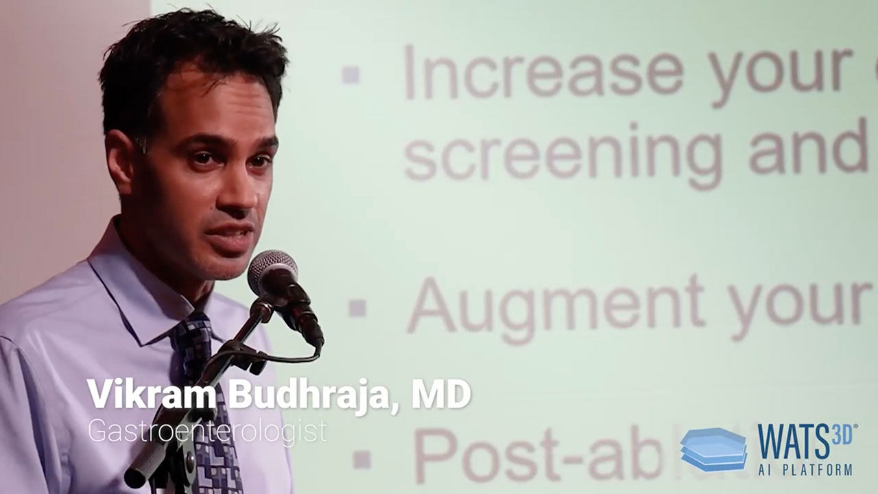
AI-Powered Analysis Enables You To Confidently Find More BE & Dysplasia
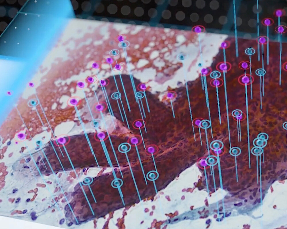
WATS3D utilizes a powerful combination of enhanced wide area sampling, 3D imaging with AI neural network analysis and expert GI pathologists - resulting in more precise diagnoses.
See How WATS3D Works
Why You Should Consider Adding WATS3D To Your EGDs
Alarming Esophageal Cancer Stats

EAC incidence rise is almost unchanged in the United States, resulting in a 700+% increase over the past 40 years. Esophageal cancer continues to be one of the deadliest (~80% 5-year mortality rate).
Limitations of Current Standard

Seattle protocol lacks utility and confidence, being time-consuming with low physician adherence. Random sampling covers only a fraction of the esophagus, leaving ~94% uncovered, prone to sampling error.
- Subtle appearance of dysplasia in BE mucosa makes dysplasia challenging to identify.
- Only 5-10% of the entire BE mucosa is sampled.
- Decrease in dysplasia detection due to poor adherance to surveillance guidelines.
- High interobserver variability due to challenges in histologic diagnosis of dysplasia.
.
Effective Common Sense Diagnostic Solution

3-tiered approach with enhanced wide-area tissue acquisition and AI analysis overcomes sampling error, reduces pathological misses, and empowers WATS3D GI pathologists to render a more precise diagnosis.
ASGE Practice Guideline:
“In this document, the ASGE offers evidence-based clinical practice guidelines on topics regarding screening and surveillance for BE.” “In patients with known or suspected BE, we suggest using WATS3D in addition to WLE with Seattle protocol biopsy sampling compared with WLE with Seattle protocol biopsy sampling alone.”
“Wide-area trans-epithelial sampling with three-dimensional computer-assisted analysis (WATS 3D , CDx Diagnostics) is a safe and effective adjunct to forcep biopsies in the evaluation of Barrett’s esophagus, LGD, and HGD." SAGES TAVAC
“The American Foregut Society (AFS) Board has concluded that there are sufficient data to support the routine use of WATS3D technology in the diagnosis and ongoing evaluation of Barrett’s esophagus.”
- Demonstrated clinical utility, effective for screening, surveillance and post-ablation - regardless of segment length.
- Evidence-based - 20+ published studies