wats3d-logo-dark

AI-powered diagnostic platform to help prevent esophageal cancer.

Included in the Standard of Practice Guidelines for the screening and surveillance of Barrett’s esophagus, WATS3D is our proprietary diagnostic platform for identifying precancerous cells in the esophagus.

AI-neural2

SEE HOW WATS3D WORKS

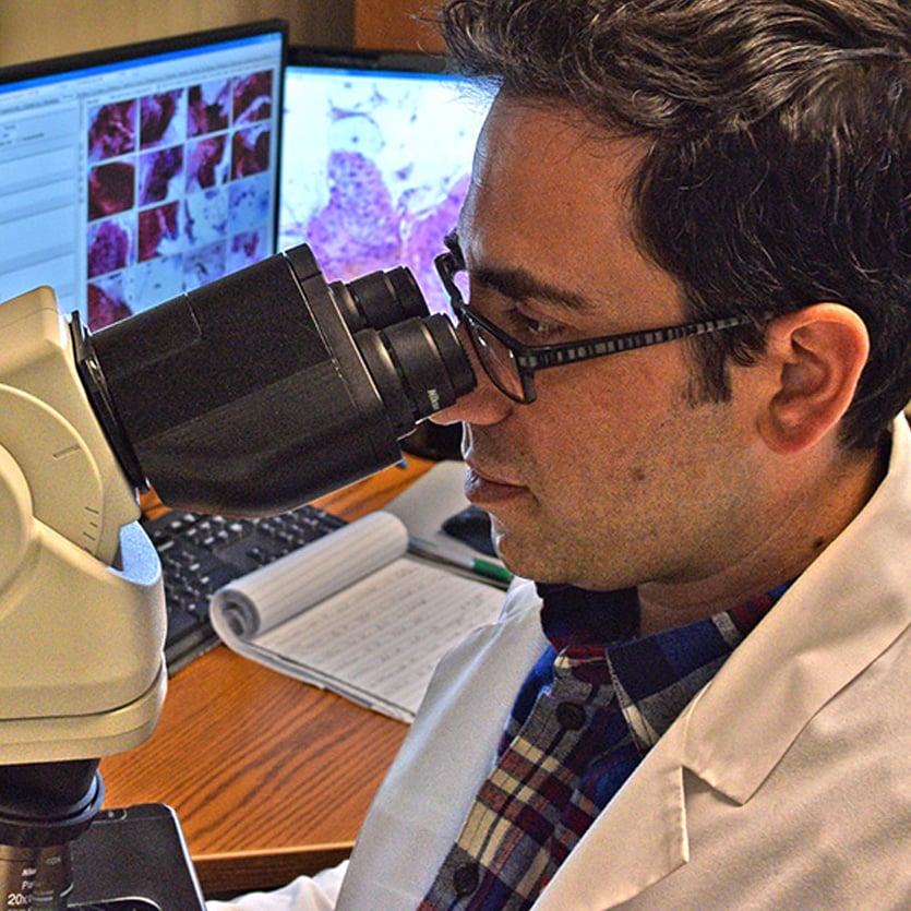
Learn how 3-Dimensional imaging with AI analysis aids pathologists in the reliable diagnosis of BE and dysplasia (3:02)

Technology

WATS3D empowers physicians to preempt cancer.

Changing the landscape of cancer prevention through innovative technology solutions.

Adjunctive use of WATS3D helps overcome sampling error that is inherent in the Seattle protocol

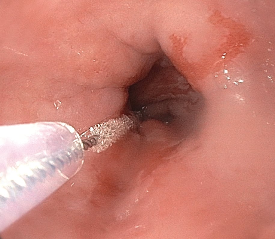
WATS3D provides physicians with a more effective endoscopic method of sampling tissue from the esophagus. It allows:

  • Sampling of much more at-risk mucosa, which reduces sampling error
  • Acquisition of deep mucosal tissue which is the location where pre-cancer (dysplasia) develops initially.


Limitations of the Seattle protocol:

  • 96% of the endoscopically suspect area remains completely unsampled
  • Significant sampling error and false negative results
  • Poor agreement rate among pathologists in diagnosing dysplasia
  • Time-consuming and labor intensive

"An estimated 33% of esophageal adenocarcinomas were diagnosed within 1 year of negative index endoscopy.”

— David A. Johnson, MD reviewing Visrodia K et al. Gastroenterology 2016 Mar

Clinical Data

WATS3D increased the overall detection of esophageal dysplasia by 242% and Barrett’s by 153%.

Adjunct use of our diagnostic platform has been vigorously tested for effectiveness in a clinical setting. Our largest study to date, involving more than 12,000 patients, yielded the results above. To put it simply: WATS3D has demonstrated reliable, consistent detection of more disease than standard biopsy methods alone.

clinical data on laptop

Case Studies

Barrett's Esophagus and Low Grade Dysplasia Identified with WATS3D and Missed by Forceps Biopsy

68 year-old female with a chronic history of achalasia that had been treated with pneumatic dilation. Patient had been well managed for decades but developed persistent reflux symptoms. Forceps biopsy results were positive for Barrett’s metaplasia without dysplasia. WATS3D results demonstrated columnar epithelium with goblet cell metaplasia (Barrett's esophagus) with low-grade dysplasia involving the crypt.

See Case Study

MatthewMcKinleyMD
"We need to make sure we use a procedure that’s accurate, safe, efficient, and cost effective: For my practice, WATS3D checks all the boxes.”

Matthew McKinley, MD
Chairman, Department of Digestive Services, ProHEALTH Care Associates, New York

pathology partnership icon

Ready to integrate WATS3D into your practice?

EDG medical procedure with medical staff

Education & Training

Integrate WATS3D into your practice.

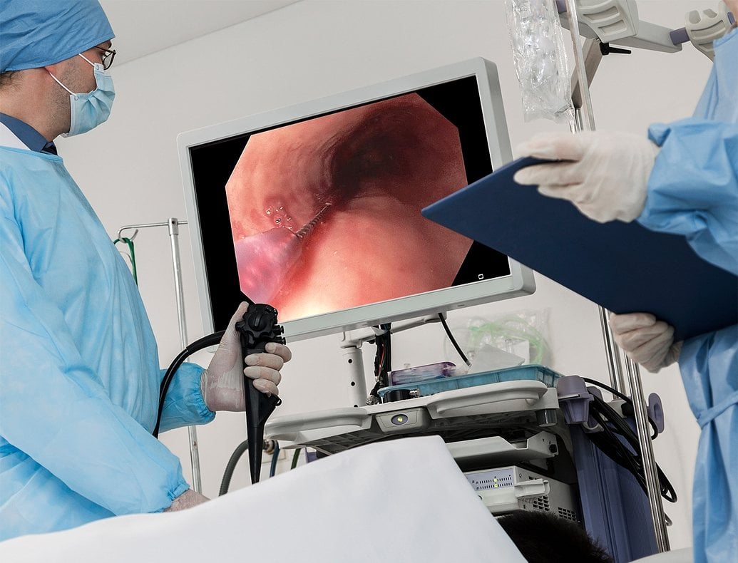
Ready to make your next EGD far more reliable? See how easy it is to implement WATS3D, the standard of care for the screening and surveillance of Barrett's esophagus, into your practice?

  • Hands-on Learning
  • Online Training
  • Educational Webinars

Learn More about Education & Training

Frequently Asked Questions about WATS3D

What are the components of the WATS3D test kit?

Each WATS3D kit contains:

  • 2 WATS3D Brush Instruments
  • 2 Vials with solution
    • 1 short Vial
    • 1 tall Vial

What size segment can be sampled with a single kit?

Each WATS3D instrument can sample a 5 cm segment.

For patients with long-segment Barrett’s esophagus, a separate kit should be utilized for every 5 cm

Ready to make your next EGD far more reliable?

It's Easy to Get Started

icon-order-kits
Order Kits

Request additional testing kits or replacement supplies.

Order Now

icon-support
Support

Let us help you with questions about our products and services.

Request support

icon-lab-services
Laboratory Services

Learn about the diagnostic assessments used in evaluating specimens.

Learn More