OralCDx®
Non-Invasive Oral Brush Biopsy with AI-enabled Tissue Analysis
A routine diagnostic test that empowers doctors to help prevent oral cancer and save lives.
OralCDx allows you to painlessly test any white or red spot in the mouth to rule out the possibility that precancerous cells are present.
If dysplasia is found, the cells can typically be removed in order to stop the progression of oral cancer.
Oral cancer is rising in low risk groups.
- Oral cancer kills about as many Americans as melanoma and twice as many as cervical cancer.
- Oral cancer is rising in women, young people and non-smokers.
- At least 25% of oral cancer victims have no known risk factors.
Finding oral cancer is too late
- By the time a lesion appears suspicious for cancer, it is generally dangerous, often life-threatening.
Mortality rates have not decreased in 50 years due to late detection.


Benefits of OralCDx®?
- Easy-to-perform
- In-office procedure - no need to reschedule patient
- Painless – well-accepted by patients
- Reliable – Clinically shown to be at least as sensitive as a scalpel in ruling our precancer or cancer.
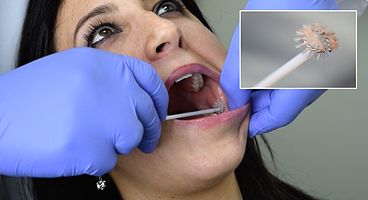
Transepithelial Sampling
- Uniquely designed sampling brush allows for the collection of cells from the full thickness of the oral epithelium.
- The disaggregated specimen requires proprietary imaging and neural network algorithm to sift through the dense tissue to find potential abnormality.
- The brush biopsy requires no anesthesia, causes no pain and minimal or no bleeding.
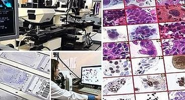
Advanced AI-Enabled
Tissue Analysis
- An advanced neural network performs tests on every cell to detect hidden abnormalities.
- The proprietary technology has been adapted for optimal detection of cellular abnormalities that are unique to oral brush biopsy samples.
- The system identifies the most suspicious cells and cell clusters and highlights them for evaluation and diagnosis by specially-trained pathologists.

Why use OralCDx®?
Due to HPV and other factors, new demographics of patients are at risk for developing oral cancer.
Any oral lesion that does not have an obvious etiology such as trauma or infection is by definition “suspicious” and requires evaluation.
Since the majority of harmless-appearing red or white spots will most likely be due to trauma, OralCDx allows you to test your patients right in your office and only refer to a specialist when it becomes clinically necessary.
OralCDx can be used to reliably rule out the possibility that an oral lesion is dysplastic or cancerous.
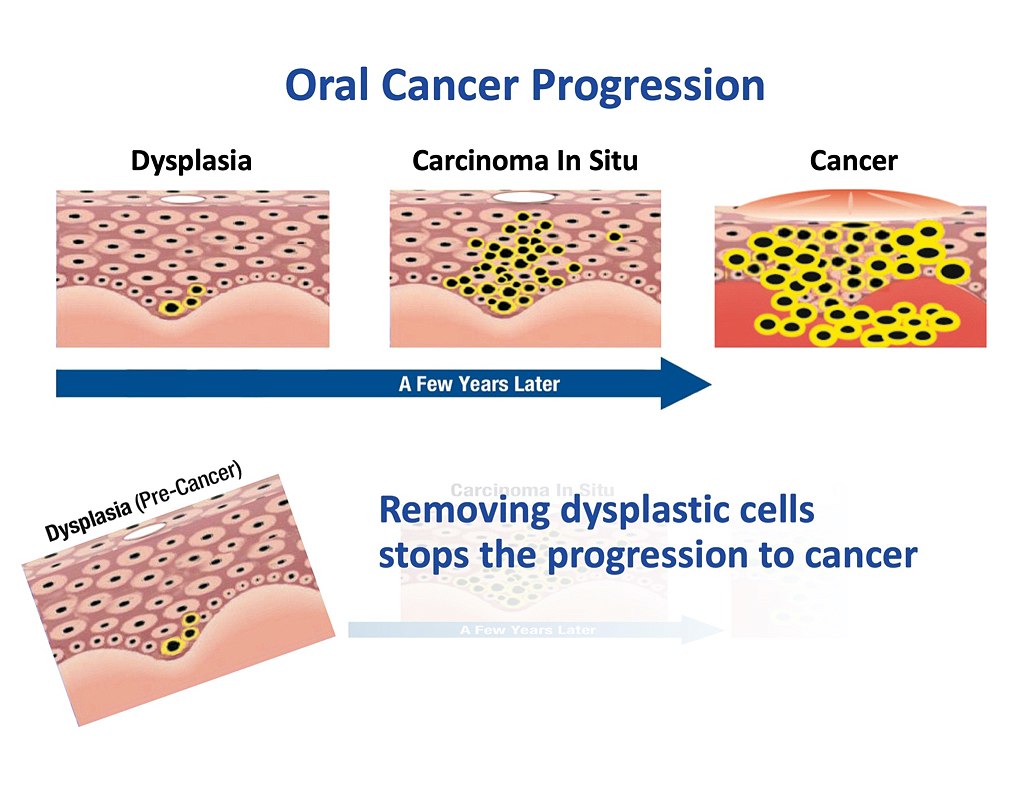
How Oral Cancer can be prevented
Virtually every oral cancer starts, years earlier, as a harmless appearing precancerous lesion. Unfortunately, precancers cannot be readily identified by their clinical features since they resemble benign, small white and red lesions that physicians encounter daily.
Just like the Pap smear is used to detect precancerous cells to help prevent cervical cancer, and colonoscopy is used to detect precancerous polyps to help prevent colon cancer, OralCDx is used to detect precancerous cells in suspicious oral lesions to help prevent oral cancer.
The progression from dysplasia to cancer occurs over a relatively long period of time, typically several years, and during this time, the lesion can be removed and oral cancer potentially prevented.
Frequently Asked Questions about OralCDx
What is the OralCDx BrushTest?
The BrushTest is a diagnostic tool that can identify pre-cancer, which is indicated by red, white or mixed spots in the mouth. The vast majority of these spots are innocuous, but 4% of them require further follow-up. If identified at this stage, oral cancer can be prevented. In the United States, more than 48,000 people are diagnosed with oral cancer annually. The disease claims the lives of nearly 10,000 a year. That’s why you should make the BrushTest an integral part of your oral cancer screening protocol.
How common are suspicious red, white or mixed spots?
They occur in about 10% of patients. If you see a suspicious spot, collect cell samples from it using the OralCDx BrushTest. Then send the collected samples to the CDx laboratory for analysis.
Of the spots you test, what percentage are precancerous?
We find abnormal cells in about 4% of cases. Many of these are dysplasia, which indicates pre-cancer.
If positive tests are so rare, why perform the test?
Because oral cancer isn’t rare. More than 48,000 people are diagnosed with oral cancer every year. By screening every patient, you can prevent oral cancer… and save lives. Since 1999, the BrushTest has detected more than 40,000 pre-cancerous spots. If a spot appears suspicious––which happens about 10% of the time–– then our recommendation is to test that spot… just to be sure that it’s not pre-cancer.
If the BrushTest detects precancerous cells, what are the next steps?
Once your office receives the results, the dentist will review the findings with the patient and then refer the patient to an oral surgeon. The surgeon will either perform a biopsy or surgically remove the precancerous spot. Most procedures are performed in-office with a local anesthesia.
Some practices use a light to screen for oral cancer. How is the BrushTest different?
The light is a visual aid, but it isn’t needed for performing an oral cancer screening. Even when a light is used, a dentist can’t tell which spots are healthy and which contain abnormal cells. Only a laboratory analysis performed by a pathologist can do that. That’s why every BrushTest includes a laboratory analysis.
Is the BrushTest accurate?
Yes. The BrushTest has been shown to be at least as sensitive as a scalpel biopsy in ruling out oral pre-cancer and cancer. Its accuracy has been demonstrated in large published studies, including a major clinical trial conducted at 35 US dental schools.
Dentists using the BrushTest have detected more than 40,000 pre-cancers since 1999.
Does the BrushTest hurt?
Most patients say that it feels like a stiff toothbrush being rubbed on the spot. Different areas of the mouth will feel different. You can use a topical anesthetic to ease any potential discomfort.
The OralCDx® brush biopsy is easy to perform.
Because of the relative ease and practicality, every oral lesion can be tested at the time it is first identified, without the need to reschedule the patient.
Obtaining cells from all the layers of the epithelium down to the basement membrane is readily accomplished by repeatedly turning the brush in a clockwise direction until the mucosa at the site of the oral lesion turns pink or red, or until pinpoint bleeding is observed.
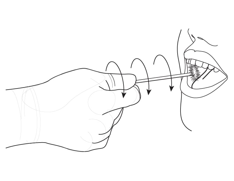
After obtaining the sample, the cellular material on the brush should be transferred to the glass slide provided in the OralCDx® kit, covered with the fixative that is supplied in individual packets and placed into the slide container supplied with the kit.
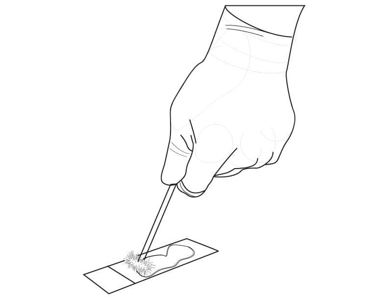
The entire test takes just a few minutes to perform and complete instructions for use are included in each kit.
If you are interested in training, please call us at 845.777.7000
View Our Latest News and Blog Posts
