Innovative technology that reliably detects dysplasia and makes cancer prevention possible.
Learn MoreStay informed about your health and the benefits of advanced technology that finds precancerous cells.
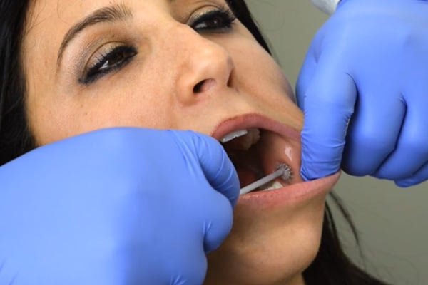
Learn MoreAll aspects of the brush have bristles. Pressure can generally be best obtained by using the flat end of the brush. However, either the flat or circular ends can be used.
Regardless of whether the flat end or side is used, ensure that you observe pinpoint bleeding.
Use of 2 brushes: Both brushes supplied in the kit should be used to test the same lesion. The first brush is rotated against the lesion until pinpoint bleeding or pinkish red tissue is observed.
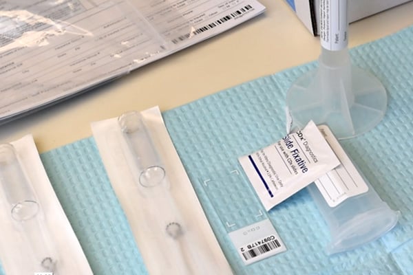
Cells are then transferred to the slide and fixed. The brush is inserted into the vial, brush side down, so that it is submerged in the liquid.
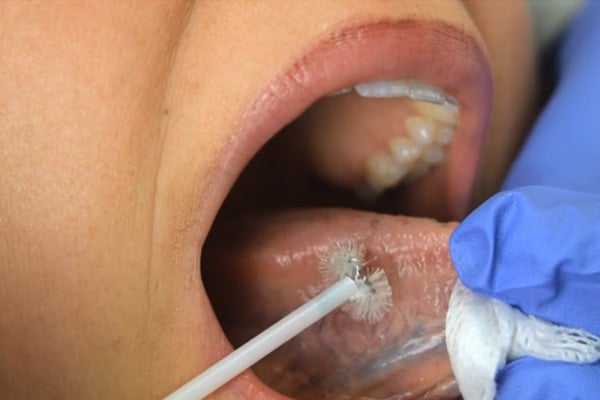
The second brush is introduced to the patient’s mouth to obtain additional cells for the cell block. After brushing, place the second brush directly into the vial (without preparation of a slide) and seal the vial tightly.
To help ensure that a full-thickness sample is obtained, rotate the brush clockwise asufficient number of times and with sufficient pressure (indicated by slight bend in brush handle) so that pinpoint bleeding occurs. That's a good indication that you have collected a full epithelial sample down to the basement membrane.
Cellular material from the brush must be transferred immediately to the slide and the fixative must be applied within 5-10 seconds. Any delay may result in air drying, which compromises the quality of the specimen.
We recommend pre-opening a fixative pack and propping it against the slide holder next to the slide before brushing so that it can be squeezed onto the slide immediately after transferring the specimen.
Additional modal content goes here. Lorem ipsum dolor sit amet, consectetur adipisicing elit, sed do eiusmod tempor incididunt ut labore et dolore magna aliqua.
©2026 CDx Diagnostics. All Rights Reserved.