EndoCDx®
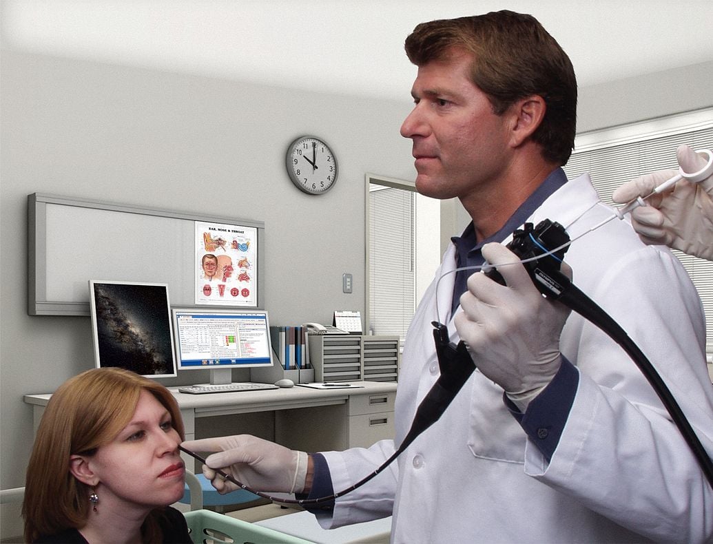
In-Office Transepithelial Laryngeal Brush Biopsy
Provides physicians with a practical method of testing non-suspicious tissue in order to help prevent laryngeal cancer.
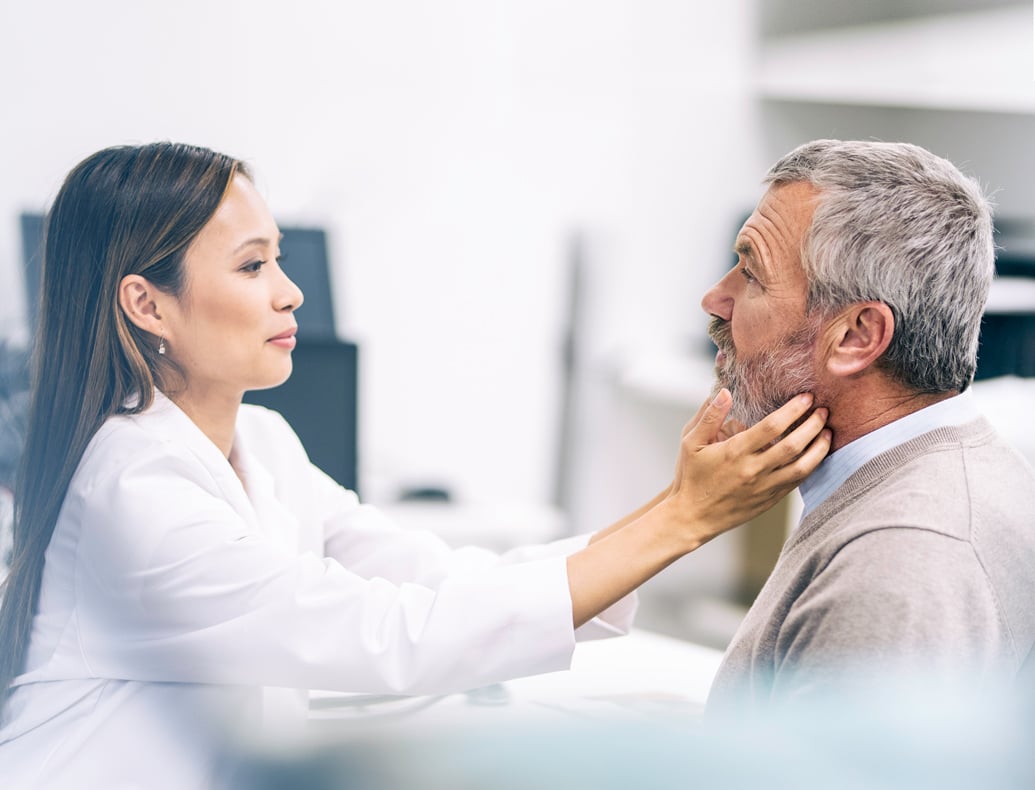
Otolaryngologists are often faced with a dilemma when relatively non-suspicious tissue is observed in the office during flexible laryngoscopy.
The probability that commonly observed tissue abnormalities are dysplastic is low, while a forceps biopsy to test these lesions presents unavoidable risks and costs. For this reason, forceps biopsy has generally been reserved for those lesions which appear relatively suspicious.
Unfortunately, by the time a lesion appears clinically suspicious, it may have already progressed to a more advanced and less easily treatable stage.
As you know, in the larynx, white or red lesions of the vocal fold and their surrounding structures are common. Repeated biopsies or excision may compromise the patient’s voice if unwanted scarring follows the biopsy. These problems are especially troublesome in preneoplastic lesions or lesions of uncertain behavior that arise from the membranous vocal folds. Repeated excisional biopsies for benign keratosis and mild reactive atypia may be avoided if these lesions can be identified and distinguished from the more aggressive lesions, such as intraepithelial neoplasia or invasive carcinoma.

Why use EndoCDx®?
EndoCDx makes obtaining a laryngeal biopsy specimen in unsedated patients possible.
- Provides practical method of evaluating innocuous areas that may have been left untested.
- Easy-to-perform
- Office-based
- Complete transepithelial sample
- Reliable AI-powered tissue pathology analysis
Laboratory Services
Browse our library of information related to esophageal cancer prevention.